Activated microglia marker
Home » » Activated microglia markerYour Activated microglia marker images are available in this site. Activated microglia marker are a topic that is being searched for and liked by netizens now. You can Download the Activated microglia marker files here. Find and Download all royalty-free vectors.
If you’re searching for activated microglia marker images information connected with to the activated microglia marker interest, you have visit the right blog. Our website frequently provides you with suggestions for seeking the highest quality video and image content, please kindly surf and find more informative video content and graphics that fit your interests.
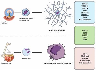
Activated Microglia Marker. Due to the shared lineage of microglia and macrophages many markers are common to both cell types. Here we directly compared the expression of HLA-DR Iba1 and CD68 in microglia with different phenotypes ranging from. HLA-DR Iba1 and CD68 are widely used. Epub 2016 Jan 1.
 Bio Bits Microglia In Inflammation And Survival From biolegend.com
Allograft inflammation factor 1 AIF-1 microglia response factor MRF-1 or daintain. However due to differences in gene regulation they may identify different activation stages of microglia. This guide presents commonly-used microglial markers with a particular focus on the new microglial-specific marker TMEM119. HLA-DR Iba1 and CD68 are widely used. In order to study the activation of microglia we established an in vitro model in which BV2 cells were cultured under hypoxia 1 oxygen. Clicking on a marker will take you to the RD Systems product selection for researching that molecule.
Ad Try sampler panel of staining validated antibodies for microglial markers.
As macrophage-like cells of the brain one of the main roles of activated microglia is that of regulating CNS innate immunity and initiating appropriate responses such. However due to differences in gene regulation they may identify different activation stages of microglia. Ad Try sampler panel of staining validated antibodies for microglial markers. This guide presents commonly-used microglial markers with a particular focus on the new microglial-specific marker TMEM119. J Cereb Blood Flow Metab. HLA-DR Iba1 and CD68 are widely used.
 Source: europepmc.org
To investigate the role of PGRN in inflammatory responses related to activated microglia we compared the immunoreactivity and expression of ionized calcium-binding adaptor molecule 1 Iba1 CD68 and CD11b as markers for activated microglia between wild-type WT and GRN-deficient KO mice. This is a member of the calcium-binding protein group. CD68 is a common marker for macrophage lineage cells primarily localized to microglia within the brain parenchyma and perivascular macrophages in. Consistent increases in markers related to activation such as major histocompatibility complex II 3643 studies and cluster of differentiation 68 1721 studies were identified relative to nonneurological aged controls whereas other common markers that stain both resting and activated microglia such as ionized calcium-binding adaptor molecule 1 1020 studies and cluster of differentiation 11b 25. J Cereb Blood Flow Metab.
 Source: researchgate.net
Ad Try sampler panel of staining validated antibodies for microglial markers. Many studies have defined phenotypes of reactive microglia the brain-resident macrophages with different antigenic markers to identify those potentially causing inflammatory damage. Analysis of microglia of CD200-deficient mice revealed a less ramified morphology with shorter processes and upregulation of CD45 leukocyte common antigen and CD11b complement receptor 3integrin α m β 2 which are also markers of activation Hoek et al 2000. CD11b CD45 Iba1 TMEM119. J Cereb Blood Flow Metab.
 Source: researchgate.net
CD11b CD45 Iba1 TMEM119. Ad Try sampler panel of staining validated antibodies for microglial markers. In order to study the activation of microglia we established an in vitro model in which BV2 cells were cultured under hypoxia 1 oxygen. Cultures were stained for CD11b microglia marker green DAPI nuclei blue tau axonal marker red and synapsin I synaptic vesicles marker. HLA-DR Iba1 and CD68 are widely used.
 Source: researchgate.net
Consistent increases in markers related to activation such as major histocompatibility complex II 3643 studies and cluster of differentiation 68 1721 studies were identified relative to nonneurological aged controls whereas other common markers that stain both resting and activated microglia such as ionized calcium-binding adaptor molecule 1 1020 studies and cluster of differentiation 11b 25. Function as a highly expressed microglia-specific marker in both mouse and human. Clicking on a marker will take you to the RD Systems product selection for researching that molecule. Epub 2016 Jan 1. Many studies have defined phenotypes of reactive microglia the brain-resident macrophages with different antigenic markers to identify those potentially causing inflammatory damage.
 Source: frontiersin.org
Due to the shared lineage of microglia and macrophages many markers are common to both cell types. Due to the shared lineage of microglia and macrophages many markers are common to both cell types. Cultures were stained for CD11b microglia marker green DAPI nuclei blue tau axonal marker red and synapsin I synaptic vesicles marker. Ad Try sampler panel of staining validated antibodies for microglial markers. HLA-DR Iba1 and CD68 are widely used microglia markers in human tissue.
 Source: pnas.org
When indicated before axons arrival to the axonal compartment a microglia cell line was cultured DIV3 and activated with LPS for 6 h DIV4. Cultures were stained for CD11b microglia marker green DAPI nuclei blue tau axonal marker red and synapsin I synaptic vesicles marker. In order to study the activation of microglia we established an in vitro model in which BV2 cells were cultured under hypoxia 1 oxygen. This is a member of the calcium-binding protein group. In this case activation of microglia through TLR4 appears to be neuroprotective in Alzheimers disease.
 Source: researchgate.net
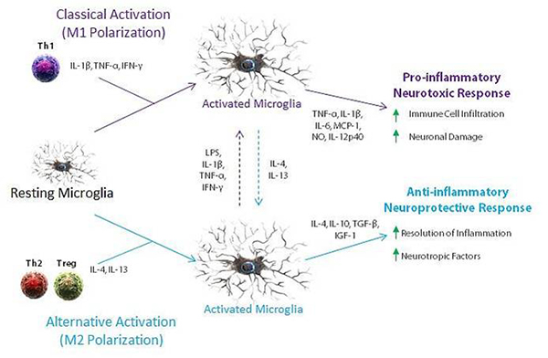
Additionally microglia in non-immunized CD200-deficient animals seemed to. To investigate the role of PGRN in inflammatory responses related to activated microglia we compared the immunoreactivity and expression of ionized calcium-binding adaptor molecule 1 Iba1 CD68 and CD11b as markers for activated microglia between wild-type WT and GRN-deficient KO mice. In contrast studies with TLR4 mutation mice indicate that microglia are activated via TLR4 signaling to reduce β-amyloid deposits and preserve cognitive functions from β-amyloid-mediated neurotoxicity. Histological characterization of the microglial phenotype revealed the elevation of classically activated microglial markers such as calgranulin B and IL-1β as well as markers associated with alternative activation of microglia including YM1 and arginase 1. It can be found under other names also.
 Source: researchgate.net
Quantitative longitudinal imaging of activated microglia as a marker of inflammation in the pilocarpine rat model of epilepsy using 11C- R-PK11195 PET and MRI. CD11b CD45 Iba1 TMEM119. This is a member of the calcium-binding protein group. However due to differences in gene regulation they may identify different activation stages of microglia. HLA-DR Iba1 and CD68 are widely used microglia markers in human tissue.
 Source: pnas.org
J Cereb Blood Flow Metab. Ad Try sampler panel of staining validated antibodies for microglial markers. Histological characterization of the microglial phenotype revealed the elevation of classically activated microglial markers such as calgranulin B and IL-1β as well as markers associated with alternative activation of microglia including YM1 and arginase 1. Due to the shared lineage of microglia and macrophages many markers are common to both cell types. Function as a highly expressed microglia-specific marker in both mouse and human.
 Source: rndsystems.com
Epub 2016 Jan 1. Once microglia encounter a substance that they sense is foreign or indicative of harm they enter an activated state. We developed monoclonal antibodies to its intracellular and extracellular domains that enable the immuno-staining of microglia in histological sections in healthy and diseased brains as well as isolation of pure nonactivated microglia by FACS. Consistent increases in markers related to activation such as major histocompatibility complex II 3643 studies and cluster of differentiation 68 1721 studies were identified relative to nonneurological aged controls whereas other common markers that stain both resting and activated microglia such as ionized calcium-binding adaptor molecule 1 1020 studies and cluster of differentiation 11b 25. This is a member of the calcium-binding protein group.
 Source: researchgate.net
Function as a highly expressed microglia-specific marker in both mouse and human. Cultures were stained for CD11b microglia marker green DAPI nuclei blue tau axonal marker red and synapsin I synaptic vesicles marker. In this case activation of microglia through TLR4 appears to be neuroprotective in Alzheimers disease. CD68 is a common marker for macrophage lineage cells primarily localized to microglia within the brain parenchyma and perivascular macrophages in. We took an alternative approach with the goal of characterizing the distribution of purinergic receptor P2RY12-positive microglia a marker previously defined as identifying homeostatic or non-activated microglia.
 Source: humanbodyworld.com
Additionally microglia in non-immunized CD200-deficient animals seemed to. Function as a highly expressed microglia-specific marker in both mouse and human. This is a member of the calcium-binding protein group. It can be found under other names also. In contrast studies with TLR4 mutation mice indicate that microglia are activated via TLR4 signaling to reduce β-amyloid deposits and preserve cognitive functions from β-amyloid-mediated neurotoxicity.
 Source: researchgate.net
Epub 2016 Jan 1. ICAM-1 was also an inflammatory factor and was reported to be produced and secreted by activated microglia which mediated the breakdown of inner blood-retinal barrier. Here we directly compared the expression of HLA-DR Iba1 and CD68 in microglia with different phenotypes ranging from. Due to the shared lineage of microglia and macrophages many markers are common to both cell types. Many studies have defined phenotypes of reactive microglia the brain-resident macrophages with different antigenic markers to identify those potentially causing inflammatory damage.
 Source: biolegend.com
Here we directly compared the expression of HLA-DR Iba1 and CD68 in microglia with different phenotypes ranging from. Once microglia encounter a substance that they sense is foreign or indicative of harm they enter an activated state. CD11b CD45 Iba1 TMEM119. HLA-DR Iba1 and CD68 are widely used microglia markers in human tissue. Microglia Activation State Markers This interactive graphic lists some of the most commonly used M1 microglia markers including iNOS COX-2 IL-6 B7-2CD86 and MHC class II molecules.
Source: journals.plos.org
Once microglia encounter a substance that they sense is foreign or indicative of harm they enter an activated state. Clicking on a marker will take you to the RD Systems product selection for researching that molecule. CD11b CD45 Iba1 TMEM119. Analysis of microglia of CD200-deficient mice revealed a less ramified morphology with shorter processes and upregulation of CD45 leukocyte common antigen and CD11b complement receptor 3integrin α m β 2 which are also markers of activation Hoek et al 2000. In order to study the activation of microglia we established an in vitro model in which BV2 cells were cultured under hypoxia 1 oxygen.
 Source: researchgate.net
Many studies have defined phenotypes of reactive microglia the brain-resident macrophages with different antigenic markers to identify those potentially causing inflammatory damage. Histological characterization of the microglial phenotype revealed the elevation of classically activated microglial markers such as calgranulin B and IL-1β as well as markers associated with alternative activation of microglia including YM1 and arginase 1. However due to differences in gene regulation they may identify different activation stages of microglia. It can be found under other names also. Additionally microglia in non-immunized CD200-deficient animals seemed to.
 Source: researchgate.net
In order to study the activation of microglia we established an in vitro model in which BV2 cells were cultured under hypoxia 1 oxygen. The most commonly used protein marker of microglia activation is an elevated level of IBA-1. Here we directly compared the expression of HLA-DR Iba1 and CD68 in microglia with different phenotypes ranging from. In order to study the activation of microglia we established an in vitro model in which BV2 cells were cultured under hypoxia 1 oxygen. CD11b CD45 Iba1 TMEM119.
Source:
Function as a highly expressed microglia-specific marker in both mouse and human. Microglia are the macrophages of the brain and spinal cord and act as an immune defense in the central nervous system CNS. Here we directly compared the expression of HLA-DR Iba1 and CD68 in microglia with different phenotypes ranging from. This is a member of the calcium-binding protein group. Consistent increases in markers related to activation such as major histocompatibility complex II 3643 studies and cluster of differentiation 68 1721 studies were identified relative to nonneurological aged controls whereas other common markers that stain both resting and activated microglia such as ionized calcium-binding adaptor molecule 1 1020 studies and cluster of differentiation 11b 25.
This site is an open community for users to do submittion their favorite wallpapers on the internet, all images or pictures in this website are for personal wallpaper use only, it is stricly prohibited to use this wallpaper for commercial purposes, if you are the author and find this image is shared without your permission, please kindly raise a DMCA report to Us.
If you find this site value, please support us by sharing this posts to your own social media accounts like Facebook, Instagram and so on or you can also bookmark this blog page with the title activated microglia marker by using Ctrl + D for devices a laptop with a Windows operating system or Command + D for laptops with an Apple operating system. If you use a smartphone, you can also use the drawer menu of the browser you are using. Whether it’s a Windows, Mac, iOS or Android operating system, you will still be able to bookmark this website.